What is the anatomy of the legs, and how can you use it to build bigger muscles? The legs are the body’s lower limbs, enabling movement while supporting the body. Therefore, we cannot easily disregard the legs and their role because they make training easy for the body. Hence, knowing about them and how to keep them in good shape must be a priority.
Every muscle in the legs, whether big or small, plays a significant role in the body. When the body rest or moves, the leg muscles work. Because most leg muscles can stretch for a distance, they call them long muscles. The small muscles assist the big muscles, maintain joints, assist in rotating joints, and enable other movements. Also, whenever the muscles contract and relax, the skeletal bones move. Regardless of their size, these muscles found in the legs are the leading causes of movement. Finally, the legs weaken when these muscles are not taken care of.
Leg Anatomy
The anatomy of the legs refers to the entire structure of the legs. This article will open your eyes to the muscles of the legs and what they do. Subsequently, the legs have four parts:
- Upper legs
- Knee
- Lower legs
- Ankle
Upper leg
This part of the legs starts from the hip to the knee and is called the thigh. The thigh is the most vital part of the lower limbs. The thigh is the most prominent part of the legs. Consequently, the thigh provides power and speed to the body. The upper portion contains the longest bone in the body. The femur bone happens to be the only bone in the upper legs. Also, it is called the thigh bone and is one of the body’s most robust bones. The femur accounts for nearly one-fourth of a person’s height. Also, it contains red bone marrow that produces most of the body’s blood.
Hamstrings
These muscles found in the upper leg allow the knee to bend. In addition, several other muscles are attached to and act on the femur. Here are the three hamstring muscles:
- Biceps femoris: This long muscle runs from the thigh region to the fibula’s head. The muscle flexes the knee and has two parts; the long head and the short head.
- Semimembranosus is a long muscle that runs from the pelvis to the tibia. This muscle helps in rotating the tibia and extending the thigh.
- Semitendinosus: The Semitendinosus is in the middle of the biceps femoris and semimembranosus. Also, this muscle helps in extending the thigh and flexes the knee.
Quadriceps
The quadriceps have four muscles that are in front of the thigh. Also, the knees can stay straightened with these muscles when bending down. Here are the four quadricep muscles:
- Vastus lateralis: Otherwise known as the vastus externus. They are the most significant and strongest muscle in the quadriceps. It helps to extend the lower legs and improve the body when standing up.
- Vastus medialis: This muscle that makes up the quadriceps starts from the thigh down to the knee. The muscle helps in extending the legs.
- Rectus femoris: It runs from the hip region down to the kneecap. The muscle helps to extend and raise the knee.
- Vastus intermedius: This muscle happens to be the deepest among the quadriceps. It acts as a cover for the femur, covering both the front and side part.
Adductors
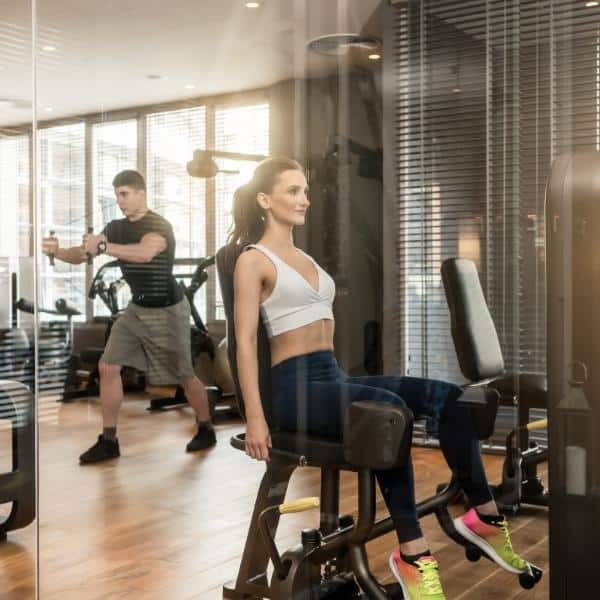
The adductors are muscles that enable the thighs to come together. They are within the thigh. Also, they play a crucial role in the anatomy of the legs and lateral movement. Finally, the abductors include:
- Adductor Magnus
- Adductor Longus
- Obturator externus
- Adductor Brevis
- Gracilis
Knee
The knee is a point in the legs where the femur, tibia, and patella meet. The patella, also known as the kneecap, works as an attachment for various tendons and ligaments. Finally, it protects the tendons and ligaments.
Tendons
The tendons are a group of tissues connected at the muscles’ end to attach the muscles to bones. The patellar tendon happens to be the largest in the knee. It attaches to the tibia and the patella. At the same time, the quadriceps tendon connects the quadriceps muscles to the patella.
Ligaments
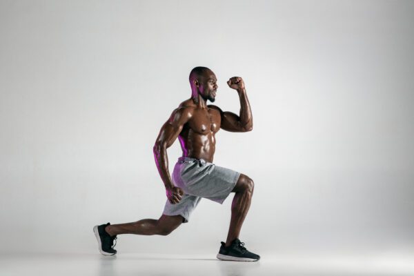
A ligament is a group of tissues connected to and surrounding a joint. Their primary duty is to support the joints and regulate the movement of the joints. Also, there are four ligaments in the knee, and they are:
- Anterior cruciate ligament: Stops the tibia in the lower leg when it wants to move too far.
- Posterior cruciate ligament: Stops the knee from moving too far backward.
- Medial collateral ligament: Helps stabilize the inner knee.
- Lateral collateral ligament: Provides stability for the outer knee.
Lower leg
The lower leg refers to the legs from the knee to the ankle, popularly called the calf. In this part of the legs, there are mainly two bones; the tibia and fibula.
- Tibia: This is larger than the fibula and is accountable for carrying weight. It is also referred to as the shinbone and is located medially.
- Fibula: The fibula is located laterally and mainly acts as a means of attachment for the lower leg muscles.
Muscles of the lower leg
- Gastrocnemius: One of the significant muscles found in the calves is the Gastrocnemius. With this muscle, a movement in the ankle called plantar flexion is possible.
- Soleus: This muscle is at the back of the Gastrocnemius and assists in plantar flexion.
- Plantaris is a minor muscle located at the lower leg’s back.
- Tibialis muscles: These muscles are on both sides of the lower leg.
- Peroneus muscles: They are on the front side of the lower leg.
Other parts of the lower leg
- Fibular nerves: These nerves stimulate muscles in the leg’s front part.
- Tibial Nerves: The tibial nerves make up the sciatic nerve. They stimulate the muscles found in the back of the lower leg.
- Achilles tendon: This tendon connects the calves’ muscles to the bones in the ankle and foot.
Ankle
What connects the lower leg to the foot is the ankle. The ankle is a joint that allows the foot’s plantar flexion and dorsiflexion. The bones that make up the ankle are the tibia, fibula, and talus. Also, in the ankle, there are two ligaments: Medial ligaments: which are often called deltoid ligaments and are in the inner ankle. Lateral ligaments: these ligaments are in the outer ankle.
Exercises for the Legs:
The anatomy of the legs can withstand high pressure and be very useful when exercising. During exercise, the muscles generate three different contractions. A concentric contraction causes muscles to shorten, thereby creating force. Eccentric contractions cause muscles to elongate in response to a greater opposing force. Isometric contractions generate power without changing the length of the muscle. Finally, the muscles in the legs work together when you perform activities or exercises. The small muscles work with the bigger muscles to get the desired results.
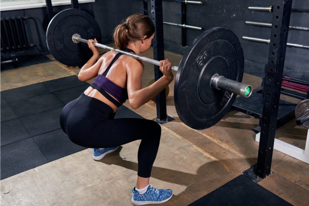
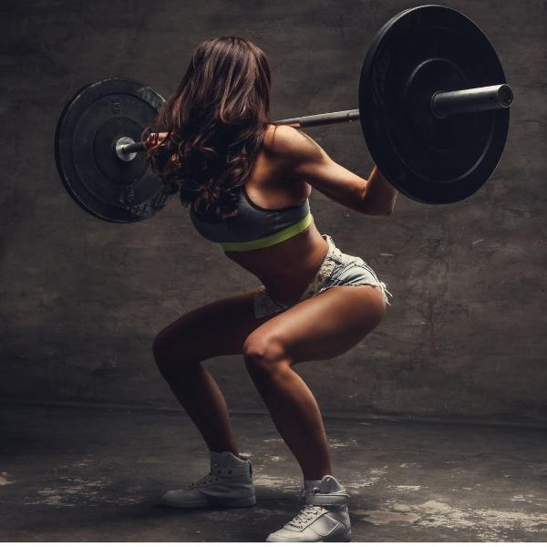
Use squats, deadlifts, leg curls, leg presses, leg extensions, and lunges to build the legs. You can use dumbbells or a barbell for standing exercises. The more weight the exercise allows you to lift, the more muscle you can build. Thus, squats and deadlifts are two of the best leg exercises.

